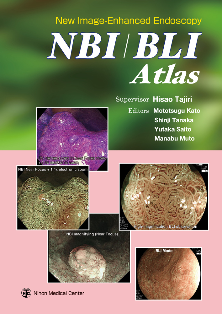
New Image-Enhanced Endoscopy NBI/BLI Atlas【電子版】

- 出版社
- 日本メディカルセンター
- 電子版ISBN
- 978-4-88875-902-1
- 電子版発売日
- 2016/12/15
- ページ数
- 246ページ
- 判型
- B5
- フォーマット
- PDF(パソコンへのダウンロード不可)
電子版販売価格:¥9,680 (本体¥8,800+税10%)
- 印刷版ISBN
- 978-4-88875-274-9
- 印刷版発行年月
- 2014/11
- ご利用方法
- ダウンロード型配信サービス(買切型)
- 同時使用端末数
- 3
- 対応OS
-
iOS最新の2世代前まで / Android最新の2世代前まで
※コンテンツの使用にあたり、専用ビューアisho.jpが必要
※Androidは、Android2世代前の端末のうち、国内キャリア経由で販売されている端末(Xperia、GALAXY、AQUOS、ARROWS、Nexusなど)にて動作確認しています - 必要メモリ容量
- 460 MB以上
- ご利用方法
- アクセス型配信サービス(買切型)
- 同時使用端末数
- 1
※インターネット経由でのWEBブラウザによるアクセス参照
※導入・利用方法の詳細はこちら
概要
目次
NBI (Narrow Band Imaging)
BLI (Blue Laser Imaging)
Oropharynx & Hypopharynx
General Theory: How to Observe These Regions with NBI or BLI
Tips on in NBI Observation
Tips on BLI Observation
Case Atlas
Case 1 NBI Inflammatory pharyngeal lesion
Case 1 NBI Inflammatory pharyngeal lesion
Case 2 BLI Hypopharyngeal papilloma
Case 3 NBI Pharyngeal melanosis
Case 4 BLI Pharyngeal melanosis
Case 5 NBI Oropharyngeal superficial carcinoma (0-IIa)
Case 6 BLI Oropharyngeal superficial carcinoma (0-IIb)
Case 7 NBI Hypopharyngeal superficial carcinoma (0-IIb)
Case 8 BLI Hypopharyngeal superficial carcinoma (0-IIa + IIb)
Esophagus
General Theory: How to Observe These Regions with NBI or BL
ITips on NBI Observation
Tips on BLI ObservationCase AtlasCase 9 NBI Glycogenic acanthosis (GA)
Case 10 BLI Esophageal papilloma
Case 11 NBI NERD
Case 12 BLI NERD
Case 13 NBI GERD
Case 14 BLI GERD
Case 15 NBI Type 0-I esophageal superficial carcinoma
Case 16 BLI Type 0-Is esophageal superficial carcinoma
Case 17 BLI Type 0-IIa esophageal superficial carcinoma
Case 18 BLI Type 0-IIb esophageal superficial carcinoma
Case 19 NBI/BLI Type 0-IIc esophageal superficial carcinoma
Case 20 NB/BLI Type 0-IIc esophageal superficial carcinoma
Case 21 BLI Type 0-IIc esophageal superficial carcinoma
Case 22 NBI Barrett's esophagus
Case 23 BLI Barrett's esophagus
Case 24 NBI Barrett's esophageal adenocarcinoma
Case 25 BLI Barrett's esophageal adenocarcinoma
Stomach & Duodenum
General Theory: How to Observe These Regions with NBI or BLI
Tips on NBI Observation
Tips on BLI ObservationCase Atlas
Stomach
Case 26 NBI Chronic gastritis
Case 27 BLI Chronic gastritis
Case 28 NBI Gastric adenoma
Case 29 BLI Gastric adenoma
Case 30 NBI Differential diagnosis of adenoma and gastric carcinoma
Case 31 BLI Differential diagnosis of erosion and early gastric carcinoma
Case 32 NBI Diagnosis of extent of early gastric carcinoma
Case 33 BLI Diagnosis of extent of early gastric carcinoma
Case 34 BLI Diagnosis of extent of early gastric carcinoma
Case 35 NBI Type 0-IIc differentiated early gastric carcinoma
Case 36 NBI Diagnosis of histological type of early gastric carcinoma
Case 37 BLI Diagnosis of histological type of early gastric carcinoma
Case 38 BLI Diagnosis of histological type of early gastric carcinoma
Case 39 NBI Transnasal endoscopic observation of early gastric carcinoma
Case 40 NBI Early carcinoma in gastric remnant
Case 41 NBI MALT lymphoma
Case 42 BLI MALT lymphoma
Duodenum
Case 43 NBI Duodenal adenoma
Case 44 BLI Duodenal adenoma
Case 45 NBI Duodenal carcinoma
Case 46 BLI Duodenal carcinoma
Case 47 BLI Duodenal carcinoma
Pathologically comparative approach to magnified images
Colon
General Theory: How to Observe These Regions with NBI or BLI
Basics of NBI Observation
Tips on NBI ObservationCase Atlas
Case 48 NBI Hyperplastic polyp
Case 49 NBI SSA/P (sessile serrated adenoma/polyp)
Case 50 BLI SSA/P (sessile serrated adenoma/polyp)
Case 51 BLI SSA/P (sessile serrated adenoma/polyp)
Case 52 NBI Elevated serrated adenoma
Case 53 NBI Elevated tubular gland duct adenoma
Case 54 NBI Superficial tubular gland duct adenoma
Case 55 NBI Tubulovillous adenoma
Case 56 BLI Villous adenoma
Case 57 NBI Elevated M carcinoma
Case 58 NBI Superficial elevated M carcinoma
Case 59 NBI Superficial depressed M carcinoma
Case 60 NBI Elevated SM carcinoma
Case 61 NBI Superficial depressed SM carcinoma
Case 62 BLI Composite (IIa + IIc) SM carcinoma
Case 63 BLI Composite (IIa + IIc) SM carcinoma
Case 64 NBI Composite (IIa + IIc) SM carcinoma
Case 65 NBI Composite (Is + IIc) SM carcinoma
Case 66 NBI LST-G, homogeneous type
Case 67 BLI LST-G, nodular mixed type
Case 68 NBI LST-NG, pseudo-depressed type
Case 69 BLI LST-NG, pseudo-depressed type
Case 70 NBI/BLI LST-NG, pseudo-depressed type
Case 71 NBI LST-NG, pseudo-depressed type
Case 72 BLI LST-NG, pseudo-depressed type
Case 73 BLI Ulcerative colitis
Case 74 NBI Tumors associated with inflammatory colon diseases (carcinoma/dysplasia)
Featured Articles
Magnifications of the Near Focus electronic zooming observation of the LUCERA ELITE system
Importance of structure enhancement in NBI magnifying observation
