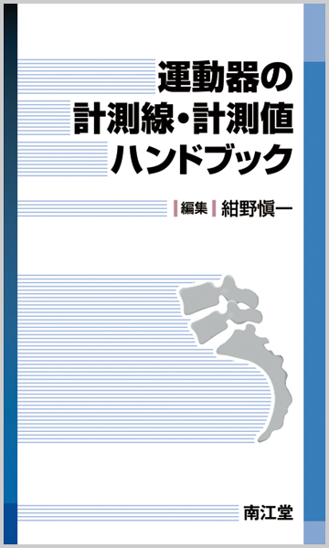
運動器の計測線・計測値ハンドブック【電子版】

- 出版社
- 南江堂
- 電子版ISBN
- 978-4-524-28601-0
- 電子版発売日
- 2016/06/06
- ページ数
- 538ページ
- 判型
- 新書
- フォーマット
- PDF(パソコンへのダウンロード不可)
電子版販売価格:¥5,500 (本体¥5,000+税10%)
- 印刷版ISBN
- 978-4-524-26336-3
- 印刷版発行年月
- 2012/11
- ご利用方法
- ダウンロード型配信サービス(買切型)
- 同時使用端末数
- 3
- 対応OS
-
iOS最新の2世代前まで / Android最新の2世代前まで
※コンテンツの使用にあたり、専用ビューアisho.jpが必要
※Androidは、Android2世代前の端末のうち、国内キャリア経由で販売されている端末(Xperia、GALAXY、AQUOS、ARROWS、Nexusなど)にて動作確認しています - 必要メモリ容量
- 40 MB以上
- ご利用方法
- アクセス型配信サービス(買切型)
- 同時使用端末数
- 1
※インターネット経由でのWEBブラウザによるアクセス参照
※導入・利用方法の詳細はこちら
この商品を買った人は、こんな商品も買っています。
概要
目次
【内容目次】
第1章 脊椎
A|脊柱
1.後弯指数|Kyphotic index
2.特発性[脊柱]側弯[症]の分類(Lenke分類)IIdiopathic scoliosis(Lenke classification)
3.側弯度(1):Cobb角|Scoliosis angle(1):Cobb’s angle
4.側弯度(2):Ferguson法|Scoliosis angle(2):Ferguson method
5.頚椎椎間可動域の測定|Range of motion of the intervertebral disc space:cervical spine
6.腰椎椎間可動域の測定|Range of motion of the intervertebral disc space:lumbar spine
B|椎体・椎間板
1.椎弓根間距離|Interpedicular distance
2.椎体[変形]指数|Vertebral body index
3.椎体楔状角|Vertebral wedge angle
4.椎間板部楔状角|Disc wedge angle
5.椎体回旋度(1):Nash & Moe法|Vertebral rotation angle(1):Nash & Moe method
6.椎体回旋度(2):Mehta法|Vertebral rotation angle(2):Mehta method
7.肋骨椎体角|Rib—vertebra angle(RVA)
8.頚椎椎間すべり距離|Gliding distance of cervical spine
9.椎間板高|Disc height index
C|頚椎
1.Chamberlain法|Chamberlain method
2.McGregor法|McGregor method
3.McRae法|McRae method
4.Bull角|Bull’s angle
5.Orbito—occipital line法|Orbito—occipital line method
6.Digastric法|Digastric method
7.歯上突起の上方移動(1):Ranawat法|Superior migration of the odontoid process(1):Ranawat method
8.歯上突起の上方移動(2):Redlund—Johnell法|Superior migration of the odontoid process(2):Redlund—Johnell method
9.後咽頭腔幅,気管後腔|Retropharyngeal space,Retrotracheal space
10.頚椎前弯|Cervical lordosis
11.石原法|Ishihara index method
12.頚椎前弯角|Cervical lordosis angle
13.C2—7角|The cervical spine angle(C2—7)
14.頚椎脊柱管前後径|Anteroposterior canal diameter of the lumbar spine
15.後縦靱帯骨化の占拠率|Occupying ratio of OPLL
16.動的因子:頚椎後方すべり|Dynamic factor(posterolisthesis of the cervical spine)
17.環椎歯突起間距離|Atlantodental distance(ADD),Atlas—dens interval(ADI)
18.脊髄余裕空間|Space available for spinal cord(SAC)
19.頚椎不安定性|Instability of cervical spine
20.棘突起間距離|Interspinous distance
21.斜台・軸椎角|Clivoaxial angle
22.延髄頚髄角|Cervicomedullary angle(CMA)
23.K—line
24.頚椎アライメント分類|Classification of cervical spine alignment
25.歯突起骨折の分類(Andersonの分類)IClassification of odontoid process fracture(Anderson classification)
26.歯突起骨形成異常の分類(Greenberg分類)IClassification of odontoid hypoplasia(Greenberg classification)
27.外傷性軸椎すべり(Levine分類)IClassification of traumatic spondylolisthesis of the axis(Levine’s classification)
28.中下位頚椎損傷分類(Allen&Ferguson分類)IClassification of lower cervical fracture and dislocation(Allen&Ferguson classification)
29.Torg—Pavlov比|Torg—Pavlov ratio
D|胸椎
1.椎弓根間距離(Elsberg—Dyke曲線)IInterpedicular distance(Elsberg—Dyke curve)
2.胸腰椎損傷(Denis分類)IThoraco—lumbar injury(Denis classification)
E|腰椎・仙骨
1.腰椎指数・腰椎インデックス|Lumbar index
2.腰椎側部指数|Lateral lumbar index(LLI)
3.腰椎脊柱管前後径|Anteroposterior canal diameter of the cervical spine
4.椎体・脊柱管比|Canal to body ratio
5.腰仙角(Dubousset法)ILumbosacral angle(Dubousset methods)
6.腰仙角|Sacral angle, lumbosacral angle, sacrohorizontal angle, sacral slope, Ferguson’s angle
7.すべり度(Marique—Taillard法)IPercent slip(Marique—Taillard method)
8.椎弓根椎間関節角|Pedicle—facet angle
9.コンパステスト|Compass test
10.腰椎分離のCT像|CT finding of lumbar spondylolysis
11.Meyerding分類|Meyerding classification
12.腰椎前弯角|Angle of lumbar lordosis
13.仙骨傾斜|Sacral inclination
14.すべり角|Slip angle
第2章 上 肢
A|上腕骨
1.上肢−上肢長管骨端間の長さ|Bone length of the upper extremities
B|肩
1.肩峰骨頭間距離|Acromiohumeral interval
2.骨頭下降率|Descending ratio of the humeral head
3.関節窩傾斜角|Glenoid tilting angle
4.Glenohumeral index
5.SCE角|Shoulder center edge angle(SCE角)
6.関節窩傾斜|Glenoid version
7.臼蓋後方開角|Posterior glenoid opening angle
8.棘上筋と棘下筋の厚み|Thickness of supraspinatus and infraspinatus
C|上腕
1.上腕骨捻転角|Humeral torsion
D|肘関節
1.キャリング角|Carrying angle
2.Baumann角(上腕骨小頭角)IBaumann angle(humerocapitellar angle)
3.上腕骨小頭傾斜角|Tilting angle(Shaft—condylar angle)
4.橈骨頭傾斜角|Radial head tilting angle
5.鉤状突起高|Coronoid height
6.肘頭鉤状突起角|Olecranon—coronoia angle
7.上腕骨角,尺骨角|Humeral(brachial)angle,Ulnar angle
8.上腕骨内旋角(山元法)IInternal rotation angle(Yamamoto method)
9.上腕骨尺骨角,上腕骨肘・手関節角|Humeral ulnar angle, Humeral elbow wrist angle
10.橈骨骨軸線|Radial shaft line
11.尺骨弯曲徴候|Maximum ulnar bow(ulnar bow sign)
12.骨幹端骨幹角|Metaphyseal—diaphyseal angle
13.前上腕線|Anterior humeral line
E|手関節・手
1.Stahl 指数|Stahl’s index
2.舟状月状骨距離(Terry—Thomas徴候)IScapholunate gap(Terry—Thomas sign)
3.橈骨月状骨角|Radiolunate(RL)angle
4.橈骨舟状骨角|Radioscaphoid(RS)angle
5.舟状月状骨角|Scapholunate(SL)angle
6.有頭月状骨角|Capitolunate(CL)angle
7.Cortical ring sign
8.掌側傾斜,背側傾斜|Palmar tilt, volar tilt:Dorsal tilt, dorsal angulation
9.橈骨傾斜|Radial inclination, radial angle
10.橈骨短縮|Radial length, shortening
11.Ulnar variance
12.尺側偏位,橈側回旋|Ulnar deviation, Radial rotation
13.Carpal height ratio
14.Carpal ulnar distance index,Carpal height ratio,Carpal—ulnar distance ratio
15.Carpal sign, Carpal angle
16.Metacarpal index(MCI)(中手骨指数)
17.Metacarpal sign(中手骨サイン)
18.Sesamoid index(種子骨指数)
19.TAM(total active motion), %TAM〔(手指の)総自動運動可動域〕
20.Pulp—distal palmar crease distance(p—p distance)(指先手掌間距離)
第3章 下 肢
A|下肢長・脚長差
1.下肢長管骨端間の距離|Length of the femur, tibia, fibula
2.下肢長管骨長|Length of the femur and tibia
B|骨盤・股関節
1.Sacro—femoral angle(仙骨大腿角)
2.Iliac angle(腸骨角)
3.臼蓋角(α角)IAcetabular index, αangle
4.臼蓋嘴(β角)IAcetabular beak angle, β angle
5.シャープ角|Sharp angle
6.臼蓋外側縁傾斜角|Slope of acetabular roof(SAR),Acetabular ridge angle(ARA)
7.Approximate acetabular index(AAI)
8.ACM角(Idelberger角)IACM angle(Idelberger’s angle)
9.臼蓋底突出距離|Protrusion distance
10.Acetabular depth
11.CE角|Center—Edge angle
12.Tear drop distance(TDD)(骨頭—涙痕間距離)
13.氏家—猪狩の外偏位角|The angle of lateral deviation(∠L)
14.山室のa値,b値|Distance a&Distance b
15.Lateral position & Superior position(Smith’s rations)
16.Acetabular—head index(AHI)(臼蓋骨頭被覆率)
17.Articulo—trochanteric distance(ATD)
18.Migration percentage(MP)
19.Hilgenreiner’s epiphyseal angle(HE angle)
20.Joint surface index(JSI),Joint surface quotient(JSQ),Radius quotient(RQ)
21.Head—neck index(HNI)
22.頚体角|Neck—shaft angle
23.Shaft—epiphysis angle
24.Neck—epiphysis angle
25.Posterior tilt angle(PTA)(後方すべり角,後方傾斜角)
26.VCA角|Vertical—center—anterior(VCA) angle
27.骨盤傾斜角(土井口)
28.Canal flare index
29.臼蓋荷重部健常域|Intact articular surface of femoral head in the weight—bearing area
30.Pelvic radius,Pelvic angle(骨盤回旋角),Pelvic morphologic angle(骨盤形態角)
31.Sacral slope,Pelvic tilt,Pelvic incidence
32.線摩耗率|Rate of linear wear
33.%Femoral offset
34.脱臼度(Crowe分類)ICrowe classification
35.応力遮蔽(Engh分類)IStress shielding(Engh classification)
36.髄腔占拠率|Canal filling ratio
37.前方開角,外方開角|Anteversion angle, Lateral opening angle
38.骨頭円形指数
39.骨頭外方化指数|Head lateralization index(HLI)
40.両側大腿骨頭中心間距離|Inter—capital distance(ICD)
41.大腿骨骨皮質インデックス|Femoral cortical index
42.前捻角|Antetosion angle,anteversion angle
43.DXA法(二重エネルギーX線吸収測定法)IDual energy X—ray absorptiometry
C|膝関節
1.膝外側角(大腿脛骨角)IFemorotibial angle(FTA)
2.下肢機能軸(Mikulicz線)IMechanical axis of the lower limb(Mikulicz line)
3.上顆軸|Epicondylar axis
4.後顆軸|Posterior condylar axis
5.前後軸|Antero—posterior axis(Whiteside’s axis)
6.The Knee Society total knee arthroplasty roentgenographic evaluation and scoring system
7.股—膝—足関節角|Hip—knee—ankle angle
8.前方ストレス撮影による垂直法・水平法
9.村瀬の中点計測法
10.野沢法
11.Heel height difference(HHD)
12.Q角|Q angle
13.顆間溝角|Sulcus angle
14.適合角|Congruence angle
15.膝蓋骨外方偏位度|Lateral shift of patella
16.膝蓋骨傾斜度|Tilting angle of patella
17.膝蓋大腿指標|Patellofemoral index
18.外側膝蓋大腿角|Lateral patellofemoral angle
19.外側膝蓋骨転位|Lateral patellar displacement
20.膝蓋骨傾斜角 IPatellar tilt
21.脛骨粗面—滑車溝間距離|Tibial tubercle—trochlear groove distance(TT—TG)
22.CTによる下肢アライメント計測|Lower limb alignment calculated by CT
23.Blumensaat線|Blumensaat’s line, Blumensaat line
24.Laurin—Labelle線|Laurin—Labelle line
25.Insall—Salvati法|Insall—Salvati method
26.Blackburne—Peel法|Blackburne—Peel method
27.Caton法|Caton method
28.Norman指数|Norman index
29.Insall—Salvati変法|Modified Insall—Salvati method
30.MRIによるpatella index(膝蓋骨指数)(Biedert)
31.Koshino—Sugimoto法|Koshino—Sugimoto method
32.骨幹端—骨幹角|Metaphyseal—diaphyseal angle
33.遠位大腿骨骨幹端角,近位脛骨骨幹端角|Distal femoral metaphyseal angle, Proximal tibial metaphyseal angle
34.脛骨内反度(脛骨内反角)IThe varus angle of the tibia
35.大腿骨内反角|The varus angle of the femur
36.Paleyの正常下肢アライメント|Normal lower limb alignment(Paley)
37.大腿外側角,脛骨外側角|Femoral angle, Tibial angle
38.反張膝角|Recurvatum angle
39.脛骨後方傾斜角|Tibial plateau angle
40.脛—腓間捻れ角|Tibio—fibular torsion
D|足関節・足
1.正面天蓋角(正面脛骨下端関節面角)IA—P mortise angle(∠TAS)
2.側面天蓋角(側面脛骨下端関節面角)ILateral mortise angle(∠TLS)
3.内果傾斜角(内果関節面角)IMedial malleolar angle(∠TMM)
4.果間角(両果下端軸角)IEmprical axis(∠TBM)
5.脛骨角,腓骨角|Tibial angle, Fibular angle
6.α角,β角Iα—angle, β—angle
7.距骨傾斜角|Talar tilt angle
8.前方引き出し距離|Anterior drawer distance
9.前方引き出し率|Anterior drawer ratio
10.脛距角|Tibiotalar angle
11.脛踵角|Tibiocalcaneal angle
12.正面距踵角|A—P talocalcaneal angle(A—P TC angle)
13.側面距踵角|Lateral talocalcaneal angle
14.横倉法(縦アーチ)IYokokura method(medial longitudinal arch)
15.踵骨傾斜角|Calcaneal pitch angle
16.側面距骨第1中足骨角|Lateral talo—first metatarsal angle(Lat—TM1,Meary’s angle)
17.踵骨第1中足骨角(Hibbs角)ICalcaneo—first metatarsal angle(Hibbs angle)
18.Bohler角|Bohler’s angle
19.足関節踵骨接床角|Ankle mortise—heel contact angle(A—H angle)
20.脛骨踵骨軸写角|Tibio—calcaneal angle(TB—C angle)
21.Steffensen&Evensen角|Steffensen&Evensen angle
22.踵部脂肪褥厚|Heel pad thickness
23.距骨軸第1中足骨基部角|Talar axis—first metatarsal base angle(TAMBA)
24.踵骨軸第1中足骨基部角|Calcaneal axis—first metatarsal base angle(CAMBA)
25.踵骨第5中足骨角|Calcaneo—fifth metatarsal angle
26.MTR角|Metatarsotalar line to the rear part of the foot angle
27.舟状骨内転度|Navicular adductor angle
28.舟状骨第1中足骨角|Naviculo—metatarsal angle(NM angle)
29.距骨長軸舟状骨横軸角|Angle between the longitudinal axis of the talus and the transvers axis of the navicular bone
30.正面距骨第1中足骨角I(A—P)Talo—first metatarsal angle(AP—TM1)
31.足軸内転度|Foot axis adductor angle
32.第2中足骨内転度I2nd metatalsar adductor angle
33.踵骨内転度|Calcaneal adductor angle
34.踵骨—両果骨|Malleolar—calcaneal angle
35.足関節前方移動量|Displacement of the ankle
36.距骨下前方移動量|Displacement of the subtalar joint
37.踵骨前方移動率 IDisplacement ratio of the subtalar joint
38.回外指数,回外角|Supination index, Supination angle
39.脛腓間距離|Distal tibio—fibular distance
40.距骨—内果間距離|Medial mortice width
41.外反母趾角|Hallux valgus angle(HV angle)
42.第1・第2中足骨間角|First—second intermetatarsal angle(M1M2 angle)
43.第1・第5中足骨間角|First—fifth intermetatarsal angle(M1M5 angle)
44.第1中足骨遠位関節面傾斜角|Distal metatarsal articular angle(DMAA)
45.趾節間外反母趾角|Hallux valgus interphalangeal angle
第4章 骨軟部腫瘍
1.砂時計腫の分類|Eden’s classification of dumbbell tumor
2.脊髄動静脈奇形|Spinal arteriovenous malformation(AVM), Spinal arteriovenous fistula(AVF)
3.長管骨病的骨折予測診断|A scoring system for diagnosing impending pathologic fractures
4.MRIで診断可能な組織や病態|Specific findings in MRI for diagnosing bone and soft tissue tumors
索引


